Going Incognito: The Invisible Universe of the Nanoplastic Pandemic
- Eugene In & Nargiss Taleb
- Jul 18, 2022
- 6 min read
Updated: Jul 30, 2022
The Plastic Pandemic
With the mass production of plastic in the 1940's, humans have come to live lives of convenience and ease. What manufacturers didn't anticipate was the vast distances across which these plastics would eventually travel — remote places such as the Balcony of Mt. Everest [1] and Antarctic ice cores [2], the digestive tracts of marine organisms [3], even in our own blood [4]. It's everywhere. Although the idea of plastic pollution has been around since the 1970's [5], it hasn't been until this year that the extensive impacts of this have been acknowledged. The beginning of March marked a historic event in which the United Nations declared by 2024 to have created an international treaty addressing the plastic problem at each stage of its lifecycle. From looking at more environmentally friendly alternatives to the management of its waste, the treaty is an awesome step in the right direction.
However, many of the mitigation steps still require us to know exactly where our plastic is in order to help regulatory communities assess their risk. Plastic in the environment is fragmented and degraded into smaller particles via various natural processes; UV-induced, thermal, and microbial processes to name a few [6]. The plastic lifecycle is long. Large (bulk) plastics degrade slowly into the well-known microplastics (akin to those of exfoliant beads in face scrubs), and eventually weathering, fragmenting over time (in various environments) into smaller nanoplastics. Plastic degradation is infinite, and has given rise to the scientific quest of detecting these seemingly ‘invisible’ plastics. Microplastics and larger fragments already have methods of detection and isolation that are long established; however, their even smaller counterparts, nanoplastics, have slipped under the radar, incognito if you will.
Only emerging in scientific literature in the last seven years, the implications of nanoplastics for human health and the environment are still riddled with uncertainties. Nanoplastics themselves are yet to have a standardised definition of their size. The issue stems from the vastly different physical and chemical properties these nano-sized materials exhibit, and by their behavior, while interacting in our macroscopic world. Understanding these materials seems unattainable (for now). While we combat the nanoplastic threat socially, the first line of active defense comes with detection. With our research, we reached our small victory in just that. Nanoplastics are invisible no more.
Before things get underway, let us introduce to you our weapon of choice, Raman Spectroscopy — the detection method that gives the possibility of pinning nanoparticle contaminants on our pollution radar.
Raman Spectroscopy
By shining a red light onto a material, you would expect the exact same colour to be reflected back. A Raman spectrometer, however, can measure that a small fraction of this reflected light is a different colour. This is a result of molecular vibrations causing a change in the energy, and therefore, the frequency of the light being scattered. As each chemical bond has its own associated energies, individual bonds (eg. C-O, C-H) and groups of bonds (e.g. benzene rings) exhibit different energy shifts and therefore, can be identified by their peak position in the Raman spectra. The benefits of Raman are the incredibly simple sample preparation and non-destructive nature of characterisation [7]. It is typically an effective technique for polymer identification — however, nanoplastics are unable to generate signals strong enough to give rise to spectral peaks. So, how can we use this vibrational technique to detect nanoplastics?

Figure 1. Polystyrene molecule.
Surface-enhanced Raman spectroscopy (SERS) is a technique introduced to overcome some of the limitations of traditional Raman spectroscopy. By using metallic nanoparticles, the electric field surrounding the metal surface is enhanced, allowing the amplification of scattering signals when interacting with the analyte.
Our Approach
As summer interns part of Professor Duncan McGillivray's Soft Matter group, we leaped into this exciting project, developing methodology for the detection of polystyrene nanoparticles. The studies involved the synthesis of spherical gold nanoparticles (AuNP) acting as our electric field enhancing material, this being used to detect polystyrene nanoplastics as our analyte of interest (Fig. 1).
To lay the foundations, a batch of ca. 20 nm AuNP’s were synthesised via an experimentally-optimised Reverse Turkevich method [8], which involves a particular ordered addition of key reagents. This method was named after the original publication by Turkevich et al. in 1951, for the synthesis of nano-sized spherical particles between the size range of 10 to 30 nm [9]. Frens in 1973 revised this method for ensuring monodispersity (uniform particle sizes) of colloidal gold nanoparticles [10]. The original Turkevich method requires a certain order of reagent addition (chloroauric acid (HAuCl4 ) to trisodium citrate (Na3 C6 H5 O7 )), with the reversed method requiring the inverse starting material addition — trisodium citrate added to the boiling gold solution. The monodispersed AuNP suspension was synthesied via reflux, giving it an overall characteristic ruby red colour (think cranberry juice).
Using this suspension, we created a system analogous to an open sandwich. The first layer or the ‘bread’ is a cellulose filter paper. The AuNP suspension once diluted was mixed together with a 1 mg mL-1 positively charged polystyrene suspension (Fig. 2), and was deposited (drop-casted) like a ‘spread’ onto the filter paper. Once dried, the monochromatic red laser (785 nm) of the Raman Spectrometer is shone through a X50 microscope lens at 50 mW power. Our vibrational spectral data was collected from a wavenumber range of 200-1800 cm-1, with a 20 s scan acquisition time. An example of the experimental set up is seen in Fig. 3, Fig. 4.

Figure 2. The system of AuNPs (-) and polystyrene (+) aggregates in a 1:1 mixture due to having opposite charges
Finding Nanoplastics — Unveiling the ‘Invisible’ Nano-world
Our results were astounding, with consistent enhancement of characteristic styrene peaks from our analyte of interest, which was positively charged 20 nm polystyrene (a common plastic). Selective polystyrene peak enhancement signals over the filter paper matrix with an AuNP capped surface was key to recording successful SERS signals.

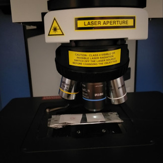
Figure 3. Inside a Raman Spectrometer lies a sample of a AuNP/ polystyrene system drop-casted onto cellulose filter paper.
With SERS effects, we can quantify the increase in signal strength via calculation of the enhancement factor (we will spare you from the math). Selective styrene peak enhancements presented enhancement factors of 110 - 1750, and in some cases, 3050 times the original signal strength were identified (Fig. 5). Excluding the 20 nm AuNP plasmon band at 518 nm, styrene related Raman signals include the aromatic C=C ring deformation (620 cm-1), C-C ring stretch (1004 cm-1), and C-H in-plane deformation (1030 cm-1). With this knowledge, we further explored the limit of detection of the filter paper SERS substrate with Raman measurements of samples with lower concentrations of polystyrene (500, 100, 50, 10, 5 and 1µg mL-1). From this we found consistent nanoplastic detection of polystyrene at concentrations of 10 µg mL-1, with instances of an all-time lowest detection limit of 5 µg mL-1 when compared to recent literature outputs.


Figure 4. A schematic illustration for the filter paper system developed in this work.
Although we had successful results in a laboratory setting, these are not necessarily representative of its native state in the environment. The effect of salt (NaCl) was tested using 150 and 600 mM concentrations, this being physiologically and seawater relevant, respectively. It was found that our ability to detect nanoplastics had reduced by a factor of 10, and our system’s limit of detection was reduced to 100 µg mL-1.
Conclusion and Future Work
Based on our filter paper-based investigation to uncover the seemingly ‘invisible’ polystyrene peaks, the developed SERS system presented a robust and efficient detection method for dilute nanoplastics in a selective manner. Consistent and reproducible styrene peak enhancements at characteristic vibrational spectroscopic stretching modes were isolated — with ability to enhance dilute concentrations of nanoplastics. Strong enhancements of positively charged polystyrene were identified, with a reliable limit of detection of 10 µg mL-1, and even as low as 5 µg mL-1. Notably, the average enhancement factor of polystyrene Raman peaks ranged from ca. 1100-1750, with an instance of enhancement performance of ca. 3050 across various AuNP batches. Our findings put to question how the interparticle distancing between the AuNP and PS spheres (mechanism) effect the enhancement factors, which can be explored using more complex methods such as small angle neutron-scattering (SANS), and small angle x-ray scattering (SAXS). To do this, a trip across the ditch to our Australian friends would be required.
Whilst we are far from reaching complex detection of nanoplastics from an environmental system with various competing matrices, our research questions the realm of nanomaterial toxicity in and around our complex, macroscopic world. Such research into the complex nanoplastic interactions are still to be pioneered. Nevertheless, a positive step forward in combatting and unveiling the ‘invisible’ plastics in simple systems offer a great potential for building on a foundation of nanoplastic detection methods, and is a small contribution (but perhaps the ultimate key) in nanotoxicology research.


Figure 5. A representative surface-enhanced Raman spectra for PS(+)20 (1.0 mg mL-1) with AuNP (orange) and non-enhanced PS(+)20 (200 mg mL-1) (black).
Acknowledgements
We were lucky enough to have jumped into this project as research interns and assistants with some amazing mentors, who we cannot thank enough for the depth of knowledge and skill development they have provided us. Thank you for teaching us to be critical and see the true art of science, and inspiring us to be better scientists and people. Thank you for the endless support and laughter, and all the banter over our many burger trips. Thank you Andrew Chan and Shinji Kihara! Absolute legends.


header.all-comments